Brain Stimulation · 2026 · Volume 19, Issue 1 · 103010
doi:10.1016/j.brs.2025.103010 · Published online 18 December 2025
Abstract
Introduction. Speech production engages a distributed network of cortical regions, but the causal dynamics within this system remain incompletely understood. Transcranial magnetic stimulation (TMS) offers a unique opportunity to modulate brain activity and assess functional contributions to behavior. However, the temporal specificity of these effects and how stimulation timing during speech impacts behavior and neural activity remains unclear.
Materials and methods. We developed a novel chronometric interleaved TMS-fMRI paradigm to deliver TMS during an overt object naming task while simultaneously recording whole-brain BOLD responses. Stimulation was applied to the left superior temporal gyrus (STG) at two task-relevant stages: during early conceptual processing and during subsequent linguistic processing. Task-related BOLD activation patterns were analyzed across conditions.
Results. TMS delivered during linguistic processing consistently induced speech arrest outside of the scanner environment and was associated with increased activation in key language areas, including the inferior frontal gyrus (IFG) and STG. In contrast, stimulation during early conceptual processing had minimal effects on neural activation. These findings suggest that stimulation-induced disruption of speech is mediated by task-state-dependent overactivation rather than suppression.
Conclusion. Our results challenge the classic “virtual lesion” view of TMS by demonstrating that stimulation can amplify neural activity in a state-dependent manner. This work establishes a novel methodological framework for temporally precise causal mapping of speech networks and emphasizes the critical role of cognitive state in shaping brain responses to stimulation.
Three key findings
- Timing matters. With the same target and pulse train, starting stimulation at 0 or 200 ms after picture onset changed the response of the language network.
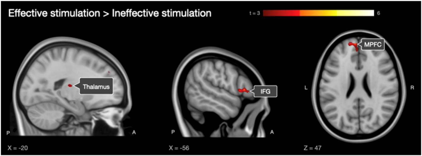
- The response extends beyond the target. In the direct comparison, later TMS onset produced greater BOLD responses in the inferior frontal gyrus, medial prefrontal cortex and thalamus.
- Speech arrest need not imply local inhibition. The findings support a state-dependent network account and challenge a purely inhibitory interpretation of TMS as a “virtual lesion”.
The study involved 20 healthy participants. Speech responses were recorded in a separate experiment outside the scanner; the TMS-fMRI experiment did not include concurrent speech recording.
Timing changes the network response
Cite this paper
Vasileiadi M, Schuler A-L, Woletz M, Witz V, Grosshagauer S, Coetzee J, Tik M. Chronometric interleaved TMS-fMRI shows state-dependent network effects underlying speech production. Brain Stimulation. 2026;19(1):103010. doi:10.1016/j.brs.2025.103010.
© 2025 The Authors · CC BY-NC-ND 4.0