Speaking requires a brain network to coordinate the meaning, sounds and movements of a word. TMS lets us interrupt that process at a chosen moment. Imaging shows how the wider network responds.
What is speech arrest?
During an object-naming task, a person sees a familiar object and says its name aloud. TMS applied to a language-relevant site can delay or interrupt that response. Researchers use these disruptions, together with other naming errors, to investigate which brain regions contribute to speech.
In our study, speech arrest and anomia during neuronavigated TMS mapping identified an individual posterior temporal target. Chronometric TMS-fMRI then tested how stimulation of that target changed activity across the language network.
From language mapping to network responses
Twenty healthy native German speakers took part in two experiments. First, a language fMRI localiser guided TMS mapping outside the scanner. Recorded naming responses identified the site most consistently associated with speech arrest or anomia. In the second experiment, participants named objects during interleaved TMS-fMRI, with stimulation beginning at either picture onset or 200 ms later.
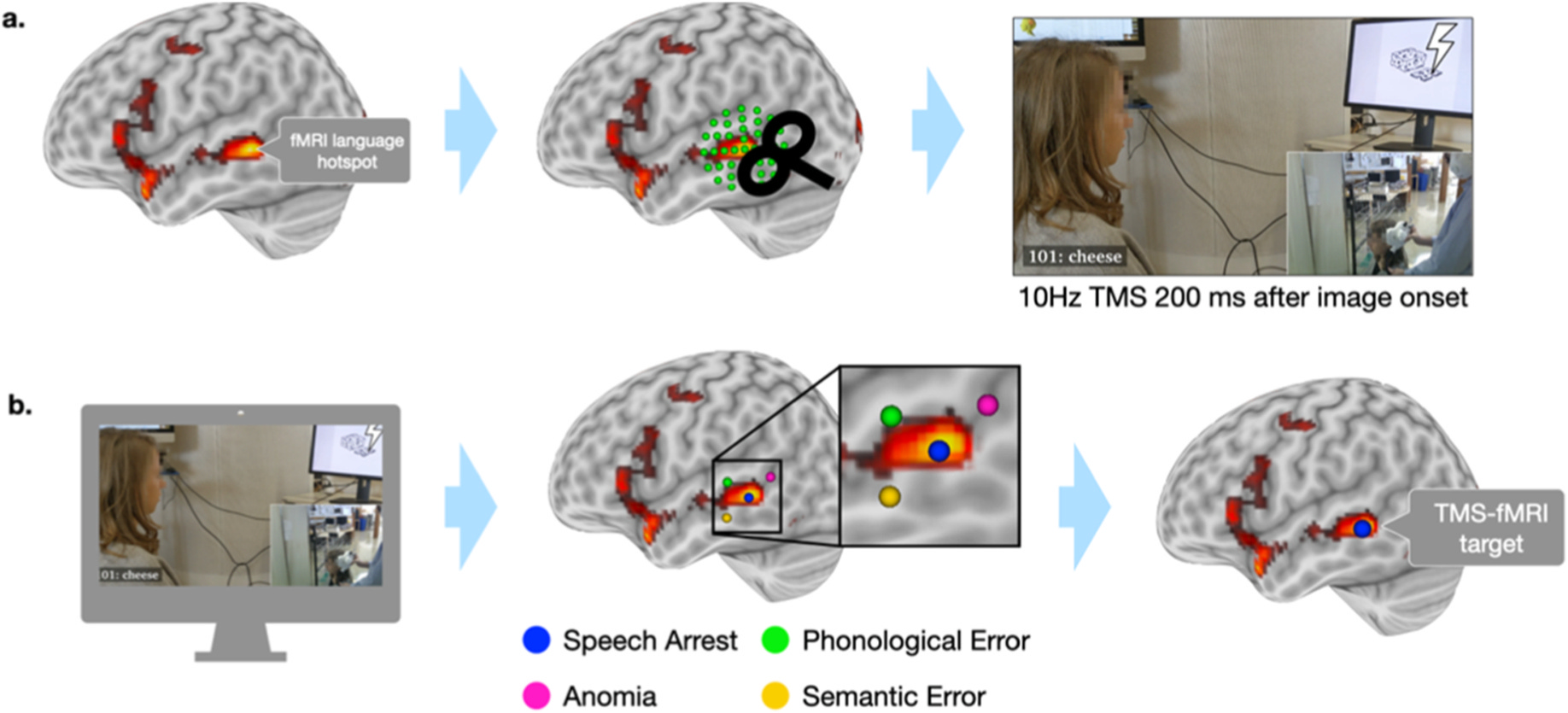
Speech errors were measured in the mapping experiment. The scanner experiment measured BOLD responses; speech output was not recorded concurrently.
Why does the stimulation time matter?
Recognising a picture, retrieving a word and preparing its pronunciation unfold over time. A perturbation therefore encounters a changing network. We kept the individual target and pulse pattern constant while shifting the onset of the TMS burst.
The 0 ms and 200 ms conditions sample different, partly overlapping processing windows. Comparing them tests how task timing changes the brain’s response to the same stimulation.
What changes across the language network?
The direct comparison showed greater BOLD responses for 200 ms than 0 ms stimulation in the inferior frontal gyrus, medial prefrontal cortex and thalamus. Stimulation of a posterior temporal target therefore changed responses beyond the stimulated region.
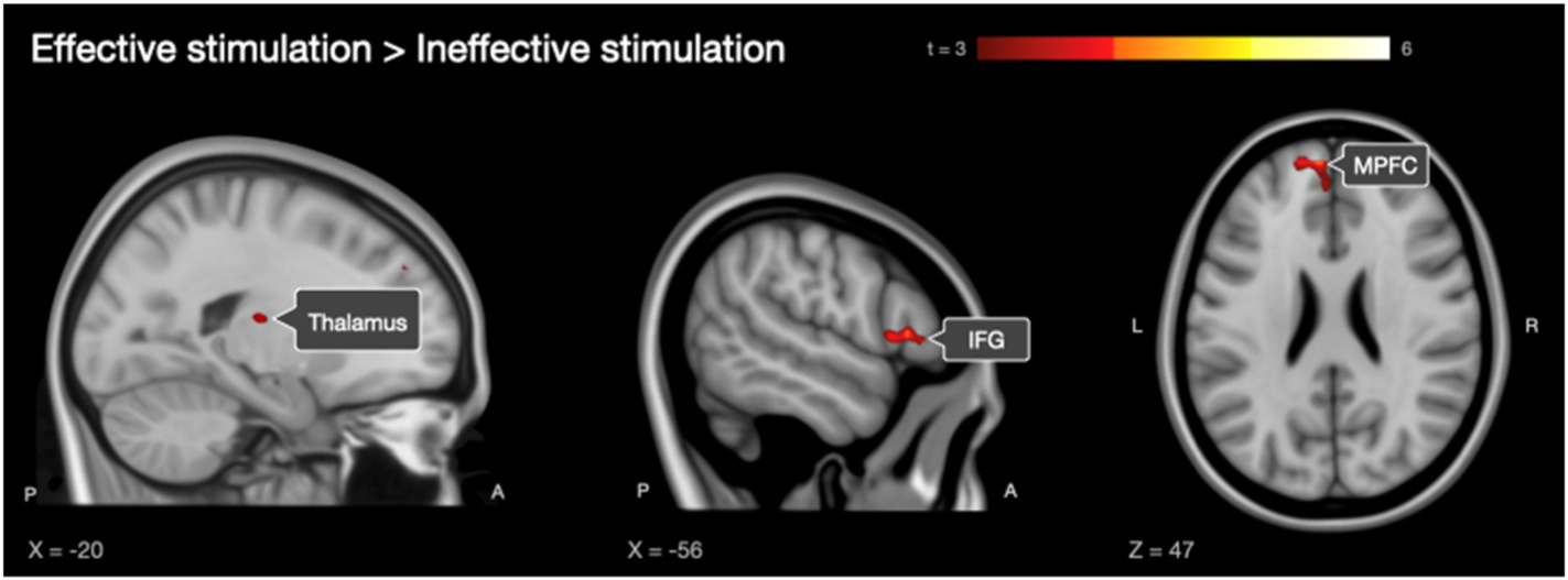
The regional pattern matters: STG responses were greater with 200 ms stimulation than with early stimulation or no stimulation. In the IFG region-of-interest analysis, 200 ms stimulation produced a greater response than 0 ms, while naming without TMS produced the highest response.
What does this mean for TMS language mapping?
A naming error identifies a disruption of speech under a particular combination of target, task, timing and stimulation parameters. Understanding that disruption also requires looking at the network in which the target participates.
Our findings support a network account of TMS-induced speech disruption: stimulation can alter activity and coordination across the language system. They challenge the assumption that a disrupted response must arise from local suppression alone. This connects language mapping with the temporal organisation of speech production and the state dependence of TMS effects.
The study
Chronometric interleaved TMS-fMRI shows state-dependent network effects underlying speech production
Maria Vasileiadi, Anna-Lisa Schuler, Michael Woletz, Verena Witz, Sarah Grosshagauer, John Coetzee and Martin Tik. Brain Stimulation, 2026;19(1):103010.